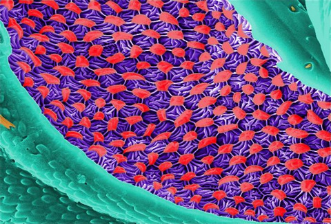
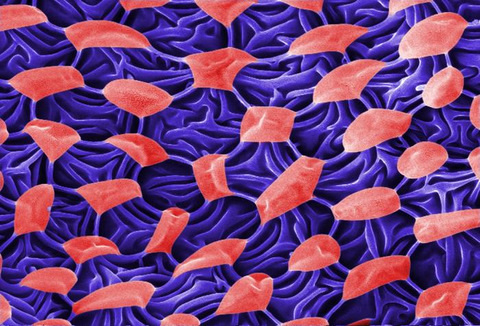
This digitally-colorized scanning electron micrograph (SEM) revealed
some of the ultrastructural morphology displayed on the ventral surface
of a bedbug, Cimex lectularius. From this view, at the top, you can see
the insect’s skin piercing mouthparts it uses to obtain its blood meal,
as well as a number of its disarticulated six jointed legs. You’ll also
notice a beautiful diaphanous structure at the bottom of the image. It
is speculated that this wondrous ultrastructural organ is most probably a
scent gland, or related to the dissemination of scent, which may be
pheromonal in nature. A further dissection of this, and the adjacent
mesothoracic region, could possibly reveal an internalized aspect of
this organ, which would be glandular in nature, and actually involved in
the production of the aromatic chemical.
Ultrastructural Morphology Displayed on the Ventral Surface of a Bedbug (Cimex lectularius)
This digitally-colorized scanning electron micrograph (SEM) revealed
some of the ultrastructural morphology displayed on the ventral surface
of a bedbug, Cimex lectularius. From this view, at the top, you can see
the insect’s skin piercing mouthparts it uses to obtain its blood meal,
as well as a number of its disarticulated six jointed legs. You’ll also
notice a beautiful diaphanous structure at the bottom of the image. It
is speculated that this wondrous ultrastructural organ is most probably a
scent gland, or related to the dissemination of scent, which may be
pheromonal in nature. A further dissection of this, and the adjacent
mesothoracic region, could possibly reveal an internalized aspect of
this organ, which would be glandular in nature, and actually involved in
the production of the aromatic chemical.
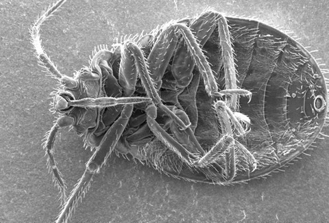
Skin Piercing Mouthparts of a Bedbug, (Cimex lectularius)
This digially-colorized scanning electron micrograph (SEM) revealed
some of the ultrastructural morphology displayed on the ventral surface
of a bedbug, Cimex lectularius. From this view you can see the insect’s
skin piercing mouthparts it uses to obtain its blood meal, as well as a
number of its six jointed legs.
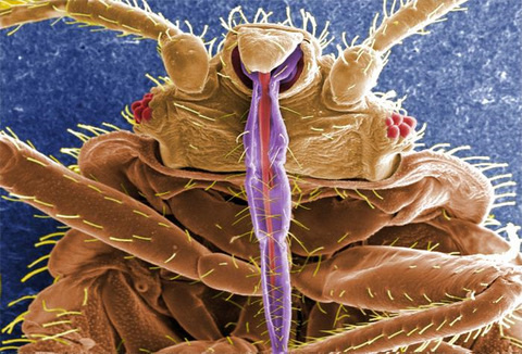
Ultrastructural Morphology on the Rostral Head Region of a Bedbug (Cimex lectularius)
This highly-magnified, digitally-colorized scanning electron
micrograph (SEM) revealed some of the ultrastructural morphology
displayed on the rostral head region of a bedbug, Cimex lectularius.
Note the proximal anatomical relationships the insect’s skin piercing
mouthparts it uses to obtain its blood meal, and how they join the head.
Ultrastructural Morphology Displayed on the Ventral Surface of a bedbug (Cimex lectularius)
This digitally-colorized scanning electron micrograph (SEM) revealed
some of the ultrastructural morphology displayed on the ventral surface
of a bedbug, Cimex lectularius. From this view, at the top, you can see
the insect’s skin piercing mouthparts it uses to obtain its blood meal,
as well as a number of its disarticulated six jointed legs. You’ll also
notice a beautiful diaphanous structure at the bottom of the image. It
is speculated that this wondrous ultrastructural organ is most probably a
scent gland, or related to the dissemination of scent, which may be
pheromonal in nature. A further dissection of this, and the adjacent
mesothoracic region, could possibly reveal an internalized aspect of
this organ, which would be glandular in nature, and actually involved in
the production of the aromatic chemical.
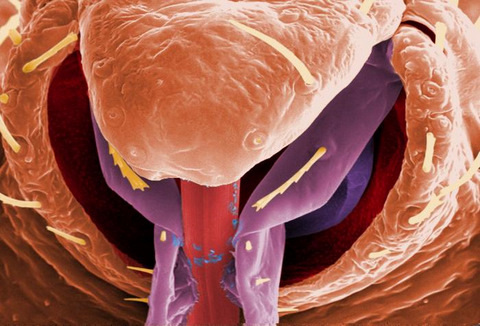
Ultrastructural Morphology on the Ventral Surface of a Bedbug (Cimex lectularius)
This scanning electron micrograph (SEM) revealed some of the
ultrastructural morphology displayed on the ventral surface of a bedbug,
Cimex lectularius. From this view you can see the insect’s skin
piercing mouthparts it uses to obtain its blood meal.
Ultrastructural Morphology on the Head of a Bedbug (Cimex lectularius)
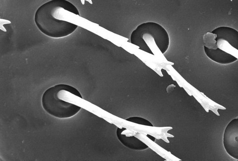
This scanning electron micrograph (SEM) revealed some of the
ultrastructural morphology displayed on the head region of a bedbug,
Cimex lectularius. In this particular view, what appeared to be “hairs”
were not hairs at all, but sensory structures known as “setae”, which
are composed of chitin, the same material as the rest of this organism’s
exoskeleton.
Chitin is a molecule made up of bound units of acetylglucosamine, joined in such a way as to allow for increased points at which hydrogen bonding can occur. In this way chitin provides increased strength, and durability as an exoskeletal foundation.
Chitin is a molecule made up of bound units of acetylglucosamine, joined in such a way as to allow for increased points at which hydrogen bonding can occur. In this way chitin provides increased strength, and durability as an exoskeletal foundation.
Dorsal View of a Bedbug Nymph (Cimex lectularius)
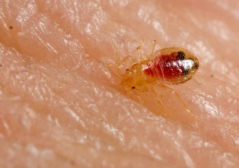
This photograph depicts a dorsal view of a bedbug nymph, Cimex
lectularius, as it was in the process of ingesting a blood meal from the
arm of a “voluntary” human host, which could be seen filling the
insect’s abdomen.
Bedbugs are not vectors in nature of any known human disease. Although some disease organisms have been recovered from bedbugs under laboratory conditions, none have been shown to be transmitted by bedbugs outside of the laboratory.
The common bedbug is found worldwide. Infestations are common in the developing world, occurring in settings of unsanitary living conditions and severe crowding. In North America and Western Europe, bedbug infestations became rare during the second half of the 20th century and have been viewed as a condition that occurs in travelers returning from developing countries. However, anecdotal reports suggest that bedbugs are increasingly common in the United States, Canada, and the United Kingdom.
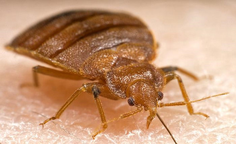
Lateral View of an Adult Bedbug (Cimex lectularius)

This photograph depicts a lateral view of an adult bedbug, Cimex
lectularius, as it is in the process of ingesting a blood meal from the
arm of a “voluntary” human host.
Bedbugs are not vectors in nature of any known human disease. Although some disease organisms have been recovered from bedbugs under laboratory conditions, none have been shown to be transmitted by bedbugs outside of the laboratory. Bedbug bites are difficult to diagnose due to the variability in bite response between people, and due to the change in skin reaction for the same person over time. It is best to collect and identify bedbugs to confirm bites. Bedbugs are responsible for loss of sleep, discomfort, disfiguring from numerous bites and occasionally bites may become infected.
Bedbugs are not vectors in nature of any known human disease. Although some disease organisms have been recovered from bedbugs under laboratory conditions, none have been shown to be transmitted by bedbugs outside of the laboratory. Bedbug bites are difficult to diagnose due to the variability in bite response between people, and due to the change in skin reaction for the same person over time. It is best to collect and identify bedbugs to confirm bites. Bedbugs are responsible for loss of sleep, discomfort, disfiguring from numerous bites and occasionally bites may become infected.
Frontal View of an Adult Bedbug (Cimex lectularius)
This photograph depicts a frontal view of an adult bedbug, Cimex
lectularius, as it is in the process of ingesting a blood meal from the
arm of a “voluntary” human host.
Bedbugs are not vectors in nature of any known human disease. Although some disease organisms have been recovered from bedbugs under laboratory conditions, none have been shown to be transmitted by bedbugs outside of the laboratory. Bedbug bites are difficult to diagnose due to the variability in bite response between people, and due to the change in skin reaction for the same person over time. It is best to collect and identify bedbugs to confirm bites. Bedbugs are responsible for loss of sleep, discomfort, disfiguring from numerous bites and occasionally bites may become infected.
Bedbugs are not vectors in nature of any known human disease. Although some disease organisms have been recovered from bedbugs under laboratory conditions, none have been shown to be transmitted by bedbugs outside of the laboratory. Bedbug bites are difficult to diagnose due to the variability in bite response between people, and due to the change in skin reaction for the same person over time. It is best to collect and identify bedbugs to confirm bites. Bedbugs are responsible for loss of sleep, discomfort, disfiguring from numerous bites and occasionally bites may become infected.
Ultrastructural Morphology on the Head of a Bedbug (Cimex lectularius)
This digitally-colorized scanning electron micrograph (SEM) revealed
some of the ultrastructural morphology displayed on the head region of a
bedbug, Cimex lectularius. Of interest is one of the insect’s compound
eyes.
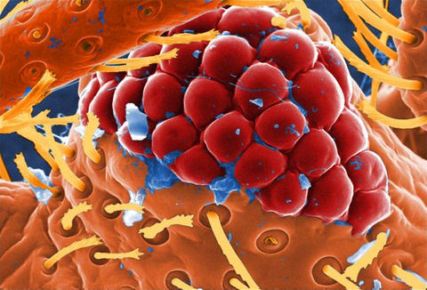
The compound eye is given this name due to the fact that the single large eye is really made up of many repeating units known as ”ommatidia”. Each ommatidium is composed of separate units made up of a photoreceptor cell, support cell, and pigment cells. Though each of these visual mechanisms functions as a separate organ, together they provide the organism with a ”compound” picture of its environment. Due to what is referred to as the ”flicker effect”, the compound eye is made very sensitive to movement, with each ommatidium turning on and off, as objects pass across its field of view. The bilateral anatomical placement of the insect’s eyes provides the organism with a very wide range of visual sensitivity.
The compound eye is given this name due to the fact that the single large eye is really made up of many repeating units known as ”ommatidia”. Each ommatidium is composed of separate units made up of a photoreceptor cell, support cell, and pigment cells. Though each of these visual mechanisms functions as a separate organ, together they provide the organism with a ”compound” picture of its environment. Due to what is referred to as the ”flicker effect”, the compound eye is made very sensitive to movement, with each ommatidium turning on and off, as objects pass across its field of view. The bilateral anatomical placement of the insect’s eyes provides the organism with a very wide range of visual sensitivity.
Tidak ada komentar:
Posting Komentar